X-Ray Microscopy of Symbolics "Ivory" CPUs.
I currently have three Symbolics MacIvory CPUs. (Do you, reader, have one to sell? Please leave a comment!)
One is currently installed in a working machine and was not yet molested; the two remaining chips, I obtained in 2017 by bartering away a Symbolics 3620.
At some point I will cut one or more of the "Ivories" open, and perform die microscopy. But prior to this, why not a bit of non-destructive imaging?
The first of the two samples is a "production" model, with a metallic cap:
Click for full resolution (1 MB):
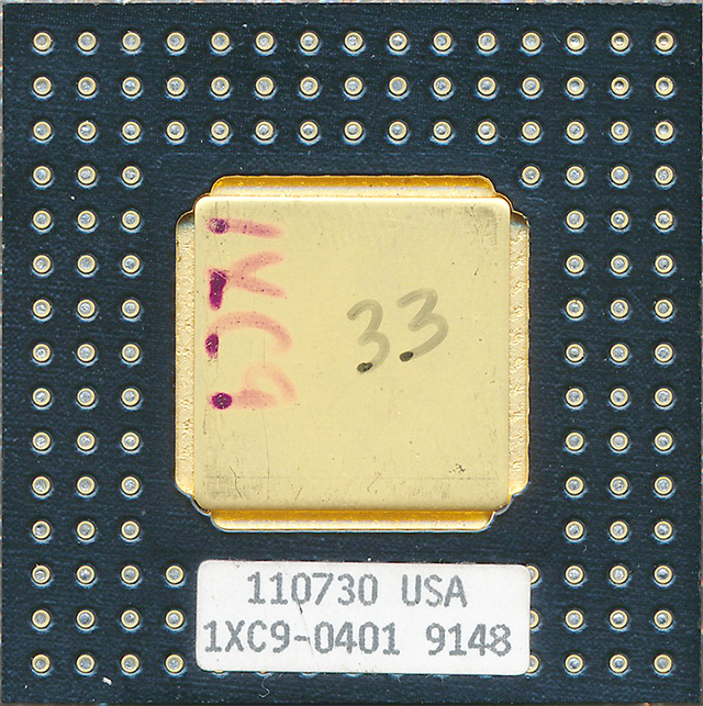
Edit: the bottom side of this unit looks like this.
An attempted die shot at maximum voltage yielded:
Click for full resolution (Warning: 20 MB!):
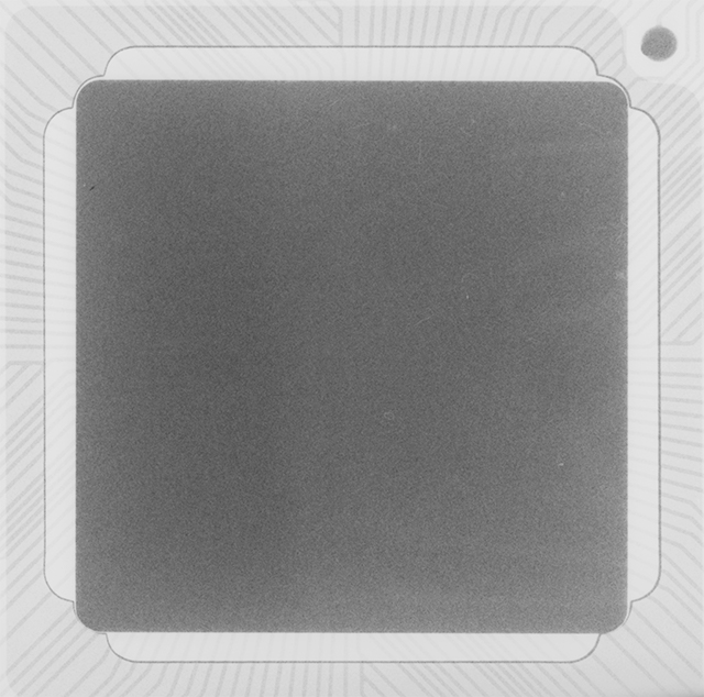
It appears that 35kV (the maximum setting of the system) is unable to produce a usefully-contrasting image of the die and connections.
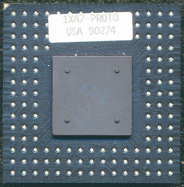
So we move on to the second sample, a "proto" model with a ceramic cap:
Click for full resolution (200 kB):
And with this one, we get:
Click for full resolution (Warning: 20 MB!):
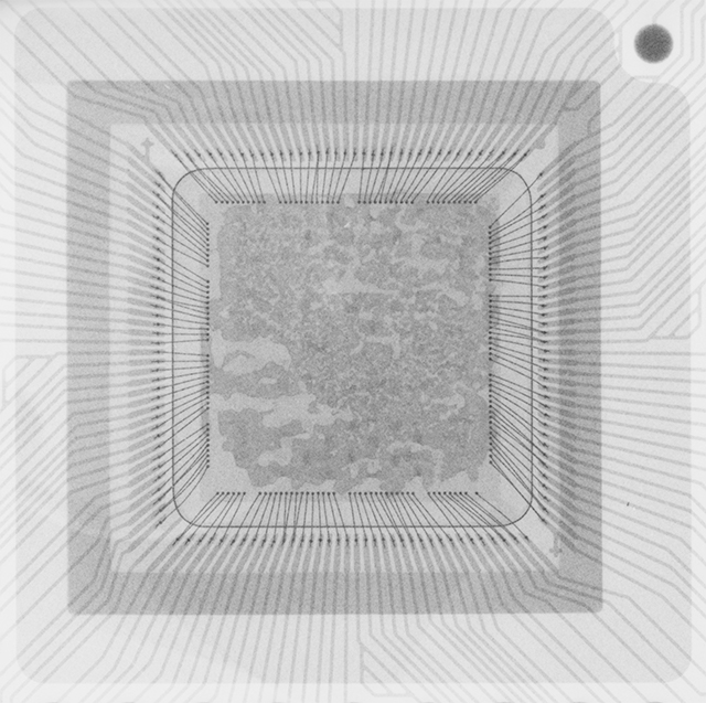
Pretty interesting. What is the substance visible on the die? (glue holding the lid in place? or a "bird's eye" view of the metallic layers of the die itself?)
Later, a magnified shot will be taken of this unit, with 35cm film and a scintillation cassette.
Exposure: 75 sec. @ 35kV, contact print, film: "Eco-30".


I don't mean to advertise but wasn't sure whether my last comment went through.
https://www.ebay.com/itm/Symbolics-Ivory-Rev-4A-CPU-chip-1XC9-0401/113755053707
Dear James,
That's DKS. Not long ago, I bought several of these from him. Shortly after this, he put some of his remaining units up on ebay, but AFAIK no takers.
If he drops the price substantially, I might consider picking up a few more.
Yours,
-S